post-title

How to Diagnose Gallbladder Stones: Tests and Procedures

Written by - Editorial Team
Medically Reviewed by - Dr Guru N Reddy

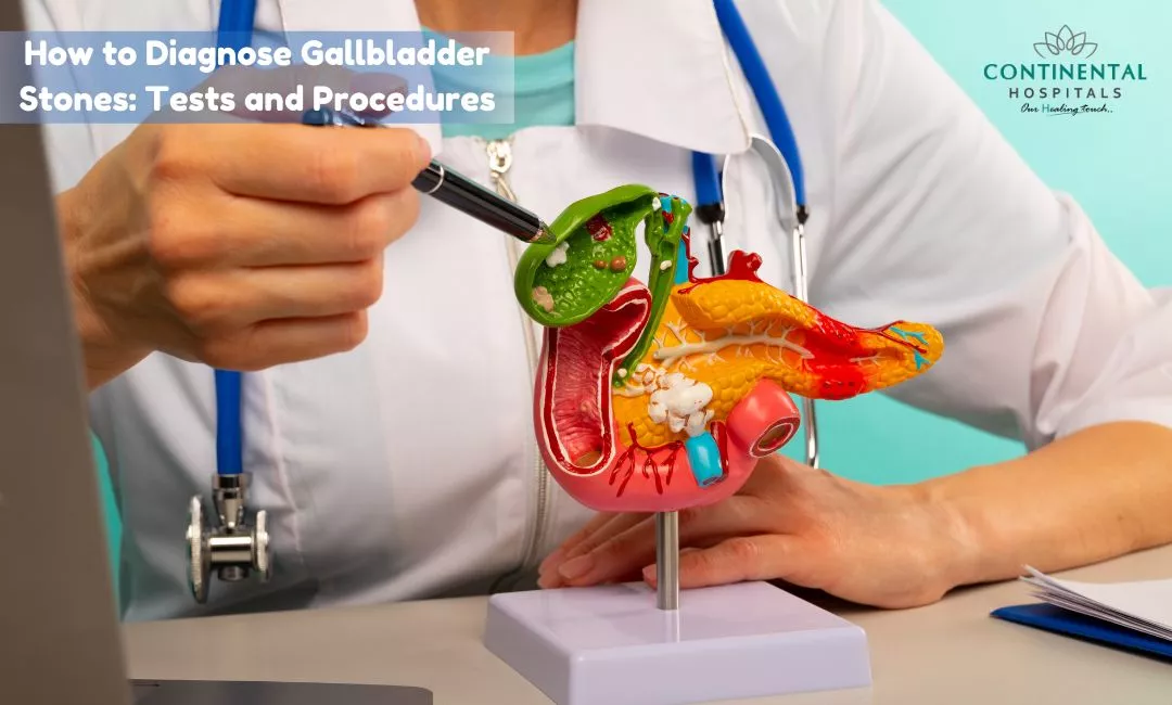
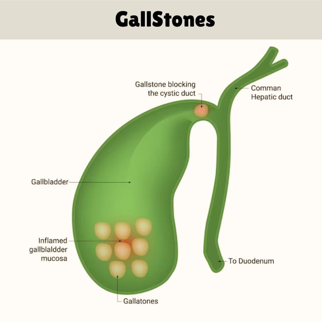
Gallbladder stones, or gallstones, are small, hard deposits that form in the gallbladder, a small organ located under the liver. They can cause pain and other symptoms, making it important to diagnose them accurately. If you’re experiencing symptoms or are concerned about gallstones, understanding the diagnostic process can help you navigate your healthcare journey more effectively. In this blog, we’ll explore the tests and procedures used to diagnose gallbladder stones and discuss treatment options available at Continental Hospitals.

What Are Gallbladder Stones?

Gallstones are solid particles that develop in the gallbladder, which stores bile produced by the liver. Bile helps digest fats, and when it becomes too concentrated, it can form stones. Gallstones come in two main types:

Cholesterol Gallstones: The most common type, formed primarily from hardened cholesterol.
Pigment Gallstones: Smaller and darker, made up of bilirubin, a substance produced from the breakdown of red blood cells.

Gallstones can vary in size and may be as small as a grain of sand or as large as a golf ball. While some people with gallstones experience no symptoms, others may suffer from pain and discomfort.

If you experience any of these symptoms, visit our gastroenterology specialist doctors for a comprehensive evaluation. Our experienced specialists provide accurate diagnosis and personalized treatment for all digestive health concerns.

What are the symptoms of gallbladder stones?

  • Abdominal Pain: Especially in the upper right side or middle of the abdomen.
  • Nausea and Vomiting: Often occurring after eating fatty foods.
  • Indigestion: Feeling bloated or gassy.
  • Jaundice: Yellowing of the skin or eyes, if the stones block bile ducts.
How are gallbladder stones diagnosed?

Diagnosing gallbladder stones typically involves a combination of medical history, physical examination, and diagnostic tests. Here’s an overview of the most common methods:

Medical History and Physical Exam

Your doctor will start by asking about your symptoms and medical history. They may also perform a physical exam, pressing on your abdomen to check for pain or tenderness. This initial assessment helps guide further testing.

Second Opinion

Ultrasound

The most common and effective test for diagnosing gallstones is an abdominal ultrasound. This non-invasive procedure uses sound waves to create images of the gallbladder and surrounding areas. Ultrasound is particularly useful because it can detect both the presence and size of gallstones and is generally safe and painless.

CT Scan

A computed tomography (CT) scan uses X-rays to create detailed cross-sectional images of the body. Although not as commonly used for gallstones as ultrasound, a CT scan can provide additional information, especially if the diagnosis is unclear or if complications are suspected.

HIDA Scan (Hepatobiliary Iminodiacetic Acid Scan)

A HIDA scan, also known as a cholescintigraphy, involves injecting a small amount of radioactive material into a vein. This tracer is absorbed by the liver and gallbladder, allowing a special camera to capture images of how the gallbladder is functioning. A HIDA scan is useful for evaluating gallbladder function and detecting conditions such as acute cholecystitis (inflammation of the gallbladder).

Endoscopic Ultrasound (EUS)

Need an Appointment?

An endoscopic ultrasound involves inserting a thin, flexible tube with an ultrasound probe down the throat and into the stomach and small intestine. This test provides detailed images of the gallbladder and bile ducts and is particularly useful for detecting small stones that may not be visible on a regular ultrasound.

ERCP (Endoscopic Retrograde Cholangiopancreatography)

ERCP is a procedure that combines endoscopy and fluoroscopy (a type of X-ray). A thin, flexible tube is inserted through the mouth and into the stomach and small intestine. A contrast dye is then injected into the bile ducts to make them visible on X-ray images. ERCP can help diagnose gallstones and is also used to remove stones or treat other complications.

Magnetic Resonance Cholangiopancreatography (MRCP)

MRCP is an MRI-based imaging technique used to visualize the bile ducts, gallbladder, and pancreatic duct. It is a non-invasive procedure that provides detailed images and can help detect gallstones, blockages, or other abnormalities. MRCP is often used when other imaging tests are inconclusive or when there is a need for a more detailed view.

Blood Tests

Blood tests may be ordered to check for signs of infection, inflammation, or liver problems. While blood tests alone cannot diagnose gallstones, they provide important information about your overall health and help identify potential complications.

What are the treatment options for gallbladder stones?

If diagnosed with gallstones, treatment options are available at Continental Hospitals, one of Hyderabad’s leading healthcare providers. The hospital offers a range of treatments to manage and treat gallbladder stones effectively:

  • Medication: For some patients, medications can help dissolve cholesterol gallstones, though this is less common.
  • Surgery: The most common treatment for symptomatic gallstones is cholecystectomy, a surgical procedure to remove the gallbladder. Continental Hospitals provides minimally invasive laparoscopic surgery, which offers faster recovery times and less post-operative discomfort.
  • Lifestyle and Dietary Changes: In some cases, dietary adjustments and lifestyle changes may be recommended to manage symptoms and prevent further stone formation.

Conclusion

Diagnosing gallbladder stones involves a combination of medical history, physical examination, and various diagnostic tests. Each test has its own strengths and can provide valuable information about the presence, size, and impact of gallstones on your health. If you suspect you have gallstones or are experiencing symptoms, it’s important to consult a healthcare professional for a thorough evaluation.

If you experience these symptoms, it’s essential to consult our best gastroenterologist in hyderabad for proper diagnosis and treatment.

Related Blogs:

  1. How to Manage Pain and Discomfort from Gallbladder Stones
  2. Managing Gallstone Pain Without Panic

Frequently Asked Questions

Gallbladder stones are diagnosed using a combination of medical history, physical examination, blood tests, and imaging studies. Your doctor will ask about symptoms such as pain in the upper right abdomen, nausea, vomiting, bloating, or pain after eating fatty foods. A physical examination helps identify tenderness or signs of infection. Blood tests check for liver function abnormalities, infection, or blockage of the bile ducts. Ultrasound is the first and most commonly used imaging test because it can clearly detect gallstones. If ultrasound results are unclear, additional imaging such as CT scan, MRI, MRCP, HIDA scan, or endoscopic ultrasound may be recommended. Early diagnosis helps prevent complications such as gallbladder inflammation, bile duct blockage, pancreatitis, or severe infection.
An abdominal ultrasound is considered the best initial test for detecting gallbladder stones. It is safe, painless, radiation free, and highly accurate for identifying stones inside the gallbladder. During the procedure, a handheld device called a transducer uses sound waves to create images of the gallbladder and surrounding organs. Ultrasound can also detect gallbladder wall thickening, inflammation, and bile duct enlargement. Most gallstones are easily visible using this test. It is often the first investigation recommended for people with symptoms of gallstones. In many cases, ultrasound provides enough information to confirm the diagnosis and guide further treatment without requiring additional imaging.
Blood tests cannot directly detect gallbladder stones, but they provide valuable information about complications caused by gallstones. Doctors may order liver function tests, bilirubin levels, pancreatic enzyme tests, and a complete blood count. Elevated liver enzymes or bilirubin may suggest a blockage in the bile ducts. Increased white blood cell counts can indicate infection or inflammation of the gallbladder. Elevated pancreatic enzymes may suggest gallstone related pancreatitis. These blood tests help doctors understand the severity of the condition and determine whether emergency treatment is needed. They are usually performed alongside imaging tests to provide a complete picture of the patient's health.
MRI and Magnetic Resonance Cholangiopancreatography, commonly known as MRCP, are recommended when doctors suspect stones in the bile ducts that may not be clearly visible on ultrasound. MRCP produces highly detailed images of the liver, gallbladder, bile ducts, and pancreas without using invasive procedures. It is particularly useful for identifying blockages, narrowing of bile ducts, or hidden stones before planning treatment. Doctors may recommend MRCP if blood tests suggest bile duct obstruction or if symptoms continue despite a normal ultrasound. This imaging technique helps avoid unnecessary invasive procedures while improving diagnostic accuracy.
A HIDA scan is a specialized nuclear medicine test that evaluates how well the gallbladder and bile ducts function. During the procedure, a small amount of radioactive tracer is injected into a vein, and a special camera tracks its movement through the liver, gallbladder, and intestines. The test is commonly used when ultrasound results are inconclusive or when doctors suspect acute gallbladder inflammation without visible stones. A HIDA scan can show whether the gallbladder is emptying normally or if bile flow is blocked. It provides valuable functional information that cannot be obtained through routine ultrasound imaging.
A CT scan can detect certain gallbladder stones, but it is generally less sensitive than ultrasound for identifying most gallstones. However, CT scans are very useful for detecting complications such as gallbladder perforation, severe infection, abscess formation, pancreatitis, or other abdominal conditions that may cause similar symptoms. Doctors may recommend a CT scan when the diagnosis remains uncertain or when they need a broader evaluation of the abdomen. Although it is not usually the first choice for diagnosing uncomplicated gallstones, it plays an important role in emergency situations and complex cases.
Endoscopic ultrasound combines endoscopy with high frequency ultrasound imaging to obtain detailed pictures of the digestive tract and nearby organs. During the procedure, a thin flexible tube with an ultrasound probe is passed through the mouth into the stomach and small intestine. This allows doctors to identify very small gallstones or bile duct stones that may not appear on standard imaging tests. Endoscopic ultrasound is especially useful when MRI or ultrasound findings are uncertain. It helps doctors accurately diagnose difficult cases and determine whether further treatment, such as stone removal, is necessary.
You should see a doctor if you experience persistent pain in the upper right abdomen, especially after eating fatty meals, or if the pain spreads to the back or right shoulder. Other warning signs include nausea, vomiting, fever, chills, yellowing of the skin or eyes, dark urine, or pale colored stools. These symptoms may indicate gallstones or complications such as gallbladder infection or bile duct blockage. Prompt medical evaluation allows early diagnosis using blood tests and imaging studies. Early treatment reduces the risk of severe complications and improves recovery outcomes while helping preserve overall digestive health.
Disclaimer: The information provided in this blog is intended for general knowledge and informational purposes only, and does not constitute medical advice. Always consult with a qualified healthcare professional for any medical concerns or before making any decisions about your health.

Get in Touch With Us

Whether you are from India or abroad, we are here to help you with your medical queries. Please fill out the form below and our team will connect with you shortly.

  • ✔ Quick response from our medical experts
  • ✔ Secure data handling & privacy
  • ✔ Easy upload for reports & documents
0 / 100
Checkboxes Section


banner_image

Chat with our medical experts on WhatsApp for quick assistance and hassle-free appointment booking

Recent Posts
Language Based Image
0 / 100