.webp)
When doctors order an X-ray for knee pain or swelling, one of the findings that may appear is a radiodense lesion. These are areas that look brighter or whiter on the X-ray because something denser than normal bone or tissue is present. But what exactly does this mean? Are radiodense lesions always serious? Let’s break it down.
What are Radiodense Lesions in the Knee?
Radiodense lesions are spots that show up on knee X-rays due to abnormal deposits, calcifications, or growths. While the term may sound alarming, not all radiodense lesions are dangerous. Some are related to benign conditions, while others may point to diseases that need close monitoring or treatment.
Understanding the possible causes helps patients know what questions to ask their doctor and why further tests might be needed.
visit the top orthopedic doctors in Hyderabad at Continental Hospitals. Our team of experienced orthopedic specialists in Hyderabad uses advanced diagnostic technology and personalized treatment plans to identify the cause of your knee problem and provide the most effective care.
What are the common conditions that cause radiodense lesions in the knee?
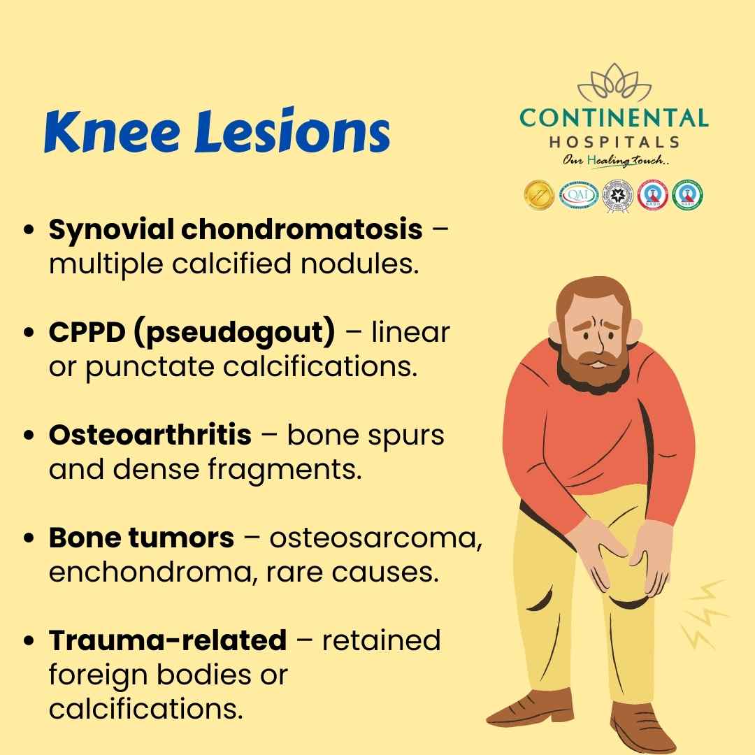
1. Synovial Chondromatosis
This is a condition where the lining of the joint (the synovium) produces small nodules of cartilage. Over time, these nodules can calcify, becoming hard and visible on X-rays as multiple white, round spots within the knee joint.
- Symptoms include pain, swelling, and sometimes stiffness.
- It often affects middle-aged adults but can appear at any age.
- Treatment may involve removing the loose bodies through minimally invasive surgery.
2. Calcium Pyrophosphate Deposition Disease (CPPD)
Commonly known as pseudogout, this condition happens when calcium pyrophosphate crystals deposit in the knee joint. On an X-ray, these crystals appear as linear or punctate calcifications within the cartilage.
- It may cause sudden, painful swelling similar to gout.
- Chronic cases can lead to joint damage and arthritis.
- Management involves medications to control inflammation and prevent flare-ups.
3. Osteoarthritis with Bone Spurs
Osteoarthritis is one of the most common reasons people get knee X-rays. As the joint wears down, the body forms bony outgrowths called osteophytes or bone spurs. These appear as dense, sharp projections around the joint edges.
- Symptoms include pain, stiffness, and reduced mobility.
- It usually develops with age or after repeated joint stress.
- Treatment ranges from physical therapy to joint replacement surgery in severe cases.
4. Tumors (Benign and Malignant)
Not all knee tumors are cancerous. Some benign bone tumors such as osteochondromas or enchondromas can create radiodense areas. However, malignant tumors like osteosarcoma may also appear dense on imaging.

- Symptoms may include persistent pain, swelling, or a lump around the knee.
- Early detection is critical, so doctors may recommend MRI or biopsy to confirm the diagnosis.
- Treatment varies depending on the type and stage of the tumor.
5. Myositis Ossificans
This condition develops when soft tissues like muscles near the knee turn into bone after injury. For example, a strong blow to the thigh or knee may trigger abnormal bone formation in the healing process. On an X-ray, this shows as a dense, irregular patch outside the normal bone.
- Common in athletes after repeated trauma.
- Usually managed with rest, physiotherapy, and sometimes surgery if severe.
6. Post-Surgical or Post-Traumatic Calcifications
After knee surgery or a significant injury, calcifications may develop in the surrounding tissues. These appear as scattered dense areas near the surgical site or fracture line.
- They may not always cause symptoms but can sometimes restrict movement.
- Follow-up imaging helps doctors decide if treatment is needed.
7. Gout
Although gout usually causes uric acid crystals, which are not radiodense themselves, long-standing gout can lead to tophi (hard uric acid deposits) that calcify and become visible on X-rays.
- Patients often experience recurrent painful flare-ups in the knee or other joints.
- Treatment involves lifestyle changes and medications that reduce uric acid levels.
8. Infections Leading to Calcifications
Chronic infections in the knee joint, although rare, can leave behind calcified areas after healing. These may appear as patchy or irregular dense spots.
- Symptoms may include persistent pain, swelling, or redness.
- Early medical attention is key to prevent joint damage.
How Do Doctors Diagnose the Cause?
An X-ray alone usually cannot provide the full picture. Doctors often combine imaging results with medical history, physical examination, and sometimes advanced tests like CT scan, MRI, or biopsy. The goal is to differentiate harmless causes from those that require urgent attention.
Key questions your doctor may ask:
- How long have you had knee pain or swelling?
- Did you have any recent injury or surgery?
- Do you have other conditions such as arthritis or metabolic diseases?
- Have you noticed lumps or worsening pain at night?
The answers, combined with imaging, guide the next steps in management.
Why Does Early Diagnosis Matter for Multiple Radiodense Lesions Around the Knee?
Ignoring knee symptoms or dismissing X-ray findings can lead to complications. For example:
- CPPD can mimic gout and cause long-term arthritis if untreated.
- Osteoarthritis may worsen over time, reducing mobility.
- Tumors, though rare, require immediate evaluation to rule out cancer.
Recognizing radiodense lesions early ensures patients get the right treatment before the condition progresses.
What are the treatment options for multiple radiodense lesions around the knee?
Treatment depends entirely on the cause:
Conservative care: Physical therapy, medications, and lifestyle changes for arthritis and crystal-related diseases.
Minimally invasive surgery: Arthroscopy to remove loose bodies in synovial chondromatosis.
Medications: Anti-inflammatory drugs, uric acid-lowering therapies, or antibiotics for infections.
Advanced interventions: Joint replacement for severe osteoarthritis or specialized cancer care for malignant tumors.
Every patient’s treatment is personalized after proper evaluation.
Why Choose Continental Hospitals for Knee Care?
Continental Hospitals is one of the leading healthcare providers with advanced imaging technology, experienced orthopedic specialists, and state-of-the-art facilities. Patients benefit from:
- Expertise in accurate diagnosis with digital imaging, MRI, and advanced pathology.
- Comprehensive care from orthopedic surgeons, rheumatologists, physiotherapists, and rehabilitation experts.
- Minimally invasive treatments that reduce recovery time and improve outcomes.
- Patient-first approach focusing on long-term knee health, not just immediate relief.
Choosing the right hospital ensures that conditions are detected early and treated effectively, giving patients the best chance for recovery and mobility.
When Should You See a Doctor?
If you notice persistent knee pain, swelling, or stiffness, it is worth getting checked. Sudden worsening of symptoms, visible lumps, or difficulty walking are clear signals to seek professional help. Remember, early diagnosis often means simpler treatment.
Conclusion
Radiodense lesions in the knee may sound worrying, but they are often linked to conditions that can be diagnosed and treated effectively with the right care. From crystal deposits and arthritis to rare tumors, understanding the possible causes helps patients make informed choices.
If you are experiencing knee pain, stiffness, or swelling, consult our Best Orthopedic Specialists in Hyderabad at Continental Hospitals. With advanced facilities and a dedicated team, you can be assured of accurate diagnosis and expert treatment to restore your knee health.