
post-title

How to Differentiate Knee Calcifications on X-Rays

Written by - Editorial Team
Medically Reviewed by - Dr Arun Reddy Mallu

Knee pain and stiffness are common complaints that bring many people to an orthopedist. Sometimes, the cause is clear, like an injury or arthritis. Other times, an X-ray reveals something unusual: white, cloudy spots in or around the joint. These are calcifications, which simply means calcium deposits in tissues that should not normally have them.

But here’s the tricky part: not all knee calcifications are the same. They can come from very different conditions, each with its own treatment approach. That is why learning how to differentiate knee calcifications on X-rays is so important for both doctors and patients. Let’s break it down in a simple way.

What are Knee Calcifications?

Calcifications are calcium buildups that appear as white, dense areas on an X-ray. Normally, calcium belongs in bones, but when it collects in soft tissues like cartilage, tendons, or joint lining, it can signal an underlying joint problem.

On X-rays, calcifications may look like:

  • Small spots or specks scattered around the joint
  • Smooth, round nodules
  • Thin, streaky or linear deposits
  • Irregular, cloudy patches

Each appearance points toward a specific condition. That’s why radiologists and orthopedic doctors pay close attention to the pattern, size, and location.

Don't ignore persistent knee pain, stiffness, or unexpected X-ray findings. Visit the top orthopedic doctors in Hyderabad at Continental Hospitals for expert diagnosis, comprehensive treatment, and the right care to restore your mobility.
What are the common conditions behind knee calcifications?

1. Synovial Chondromatosis
This is a condition where the lining of the joint (synovium) produces small nodules of cartilage, which later calcify. On X-ray, these look like multiple smooth, round calcified bodies floating inside the joint. Patients usually have pain, swelling, and limited motion.

2. Calcium Pyrophosphate Deposition Disease (CPPD)
Also known as pseudogout, CPPD happens when crystals of calcium pyrophosphate build up in cartilage. On X-rays, these appear as thin, white lines inside the joint cartilage, especially in the meniscus or articular cartilage. It often mimics osteoarthritis but may flare up suddenly like gout.

Second Opinion

3. Osteoarthritis with Calcifications
In long-standing osteoarthritis, calcium can sometimes deposit around damaged cartilage and bone spurs. The X-ray shows narrowed joint space, irregular bone surfaces, and scattered calcifications around the joint edges.

4. Gout
Although gout is primarily diagnosed by uric acid levels and symptoms, chronic gout may show calcified tophi on X-rays. These appear as irregular, lumpy calcifications around the soft tissues near the joint.

5. Tumoral Calcinosis
A rare condition, but when it occurs, it shows up as large, lobulated calcified masses around the knee joint. Unlike other causes, these deposits are usually outside the joint capsule.

6. Post-Traumatic or Post-Surgical Calcification
After an injury or surgery, calcium deposits can form in soft tissues like muscles and ligaments. These are called myositis ossificans. On X-rays, they appear as irregular, cloudy patches that may later mature into bone-like structures.

7. Retained Foreign Bodies (like acupuncture needles or fragments)
Sometimes, metallic fragments or foreign bodies show up as bright, linear densities. These can be mistaken for calcifications but have a distinct shape and density.

How do doctors differentiate knee calcifications?

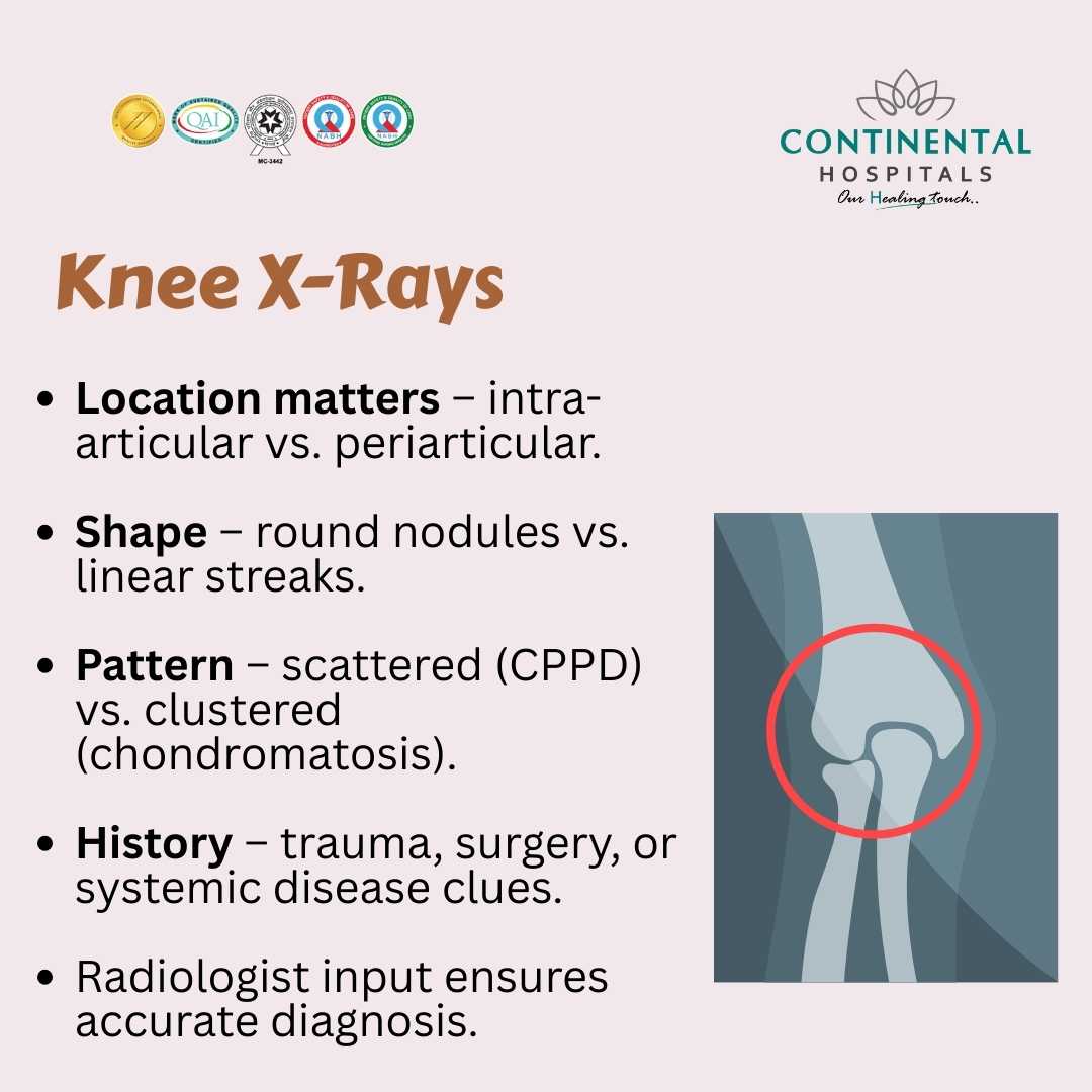
When an X-ray shows calcifications, the doctor does not stop at just looking at them. Several factors are studied:

  • Location: Are the calcifications inside cartilage, outside the joint, or within soft tissues?
  • Shape and Pattern: Smooth and round? Thin and linear? Irregular and cloudy?
  • Number: Single deposit or multiple scattered spots?
  • Associated Findings: Is there joint space narrowing, swelling, or bone damage?
  • Patient History: Has the patient had past injuries, surgeries, or crystal-related arthritis?

By combining these clues, doctors can narrow down the possible causes and decide whether additional tests like CT scan, MRI, or joint fluid analysis are needed.

Need an Appointment?

Why is it important to differentiate knee calcifications on X-rays?

Not all calcifications need treatment, but some conditions demand quick action. For example:

  • CPPD may require medicines to control pain and inflammation.
  • Synovial chondromatosis often needs surgical removal of loose calcified bodies.
  • Post-traumatic calcifications may settle with time but sometimes need intervention.
  • Gout needs long-term management of uric acid levels.

If calcifications are mistaken for something harmless, the real disease may silently worsen. That is why accurate diagnosis through X-ray interpretation is critical.

What are the symptoms of knee calcifications?

Not every patient with calcifications feels pain, but many experience:

  • Swelling around the joint
  • Stiffness or limited movement
  • Sudden attacks of sharp knee pain
  • Chronic aching and difficulty climbing stairs or walking
  • Warmth or redness in case of inflammatory conditions

If you have any of these symptoms, it’s best to consult an orthopedic specialist rather than ignoring them.

What are the treatment options for knee calcifications?

Treatment depends on the cause. Some possible approaches include:

  • Medication: Anti-inflammatory drugs for CPPD, gout, or arthritis-related pain
  • Lifestyle changes: Weight management, physical therapy, and joint-friendly exercises
  • Surgical options: Removing loose calcified nodules or correcting joint damage in severe cases
  • Underlying disease control: Managing uric acid in gout or metabolic factors in tumoral calcinosis

The key is individualized treatment guided by proper diagnosis.

Why choose Continental Hospitals for knee problems?

When it comes to musculoskeletal issues like knee calcifications, you need expert care backed by advanced imaging and orthopedic knowledge. Continental Hospitals brings together the following:

  • State-of-the-art radiology with digital X-rays, CT, and MRI for accurate diagnosis
  • Experienced orthopedic surgeons who specialize in joint disorders and complex knee conditions
  • Multidisciplinary approach where radiologists, rheumatologists, and physiotherapists work together for the best outcome
  • Personalized treatment plans that address the root cause, not just the symptoms
  • Focus on recovery and mobility through advanced physiotherapy and rehabilitation services

Choosing Continental Hospitals means you get clarity, confidence, and care you can trust.

When Should You See a Doctor?

If you notice persistent knee pain, swelling, or stiffness, or if you have already been told your X-ray shows calcifications, it is wise to consult an orthopedic doctor. Early diagnosis helps prevent complications and ensures the right treatment.

Conclusion

Knee calcifications on X-rays may look similar at first glance, but each type tells a different story about what is happening inside your joint. From cartilage changes in CPPD to nodules in synovial chondromatosis, careful evaluation is the key to proper treatment.

If you suffer from knee pain, stiffness, or unexplained X-ray findings, consult our Best Orthopedic Specialists in Hyderabad at Continental Hospitals. With expert diagnosis and comprehensive care, you can get back to moving freely without unnecessary worry.


Frequently Asked Questions

Knee calcifications are deposits of calcium that appear as white or dense areas on an X-ray. They can develop in cartilage, tendons, ligaments, blood vessels, or the tissues surrounding the knee joint. Some calcifications occur naturally with aging, while others result from injury, inflammation, arthritis, or metabolic disorders. Their appearance, location, and pattern help doctors determine the underlying cause. Small calcifications may not cause any symptoms and are often found incidentally during imaging. Larger or strategically located deposits can contribute to pain, stiffness, swelling, or reduced mobility. X-rays are the first imaging test used to identify these deposits because calcium is clearly visible. However, additional tests such as ultrasound, MRI, CT scans, or blood investigations may be needed to confirm the diagnosis and guide appropriate treatment.
Doctors differentiate knee calcifications by carefully evaluating their size, shape, density, and exact location on the X-ray. Calcifications within cartilage often suggest calcium pyrophosphate deposition disease, while deposits around tendons may indicate calcific tendinitis. Bone-forming lesions or loose bodies have distinct appearances that differ from soft tissue calcifications. Physicians also compare the findings with the patient's age, symptoms, history of injury, arthritis, or previous surgeries. Symmetry, distribution, and associated joint changes such as narrowing or bone spurs provide additional clues. Laboratory tests may be recommended if metabolic disorders are suspected. MRI or ultrasound can help assess surrounding soft tissues when needed. Combining imaging findings with clinical examination allows doctors to accurately identify the cause and develop the most appropriate treatment plan.
Yes, certain knee calcifications can be associated with different forms of arthritis. Calcium pyrophosphate deposition disease commonly produces calcifications within the cartilage, a finding known as chondrocalcinosis. Osteoarthritis may also show calcified loose bodies, bone spurs, and joint space narrowing on X-rays. These imaging findings help doctors distinguish degenerative arthritis from inflammatory or crystal-related conditions. However, not every calcification means arthritis is present. Some deposits occur without significant joint damage or symptoms. A detailed medical history, physical examination, and additional imaging or laboratory tests help confirm the diagnosis. Identifying the exact pattern of calcification is important because treatment varies depending on the underlying cause rather than the calcification itself.
No, knee calcifications are not always painful. Many people have small calcium deposits that are discovered accidentally during X-rays performed for unrelated reasons. Symptoms usually depend on the size, location, and underlying cause of the calcification. Pain may occur if the deposit irritates nearby tendons, ligaments, cartilage, or surrounding soft tissues. Some patients experience stiffness, swelling, reduced range of motion, or discomfort during walking or climbing stairs. Others remain completely symptom-free for years. If pain is persistent or worsening, further evaluation is necessary to determine whether the calcification is responsible or if another knee condition is present. Early diagnosis helps ensure appropriate treatment and prevents unnecessary complications.
Several conditions can lead to calcifications around the knee joint. Common causes include calcium pyrophosphate deposition disease, osteoarthritis, previous injuries, chronic inflammation, calcific tendinitis, bursitis, and old ligament damage. Less common causes include metabolic disorders involving calcium or phosphate balance, kidney disease, hyperparathyroidism, and certain autoimmune conditions. Previous surgeries or trauma may also leave residual calcified tissue. In rare cases, tumors or infections can produce calcified lesions that require careful evaluation. Doctors assess the appearance of the calcifications along with the patient's symptoms and medical history to identify the most likely cause. Additional imaging or laboratory tests may be recommended when the diagnosis remains uncertain.
X-rays are an excellent first-line imaging tool for detecting calcium deposits because calcifications appear clearly as dense white areas. They help determine the location, size, and overall pattern of the deposits. However, X-rays cannot always identify the exact tissue involved or reveal associated soft tissue injuries. If the diagnosis remains unclear, doctors may recommend MRI, ultrasound, or CT scans for additional detail. Blood tests may also be performed to evaluate crystal disorders, inflammation, or metabolic abnormalities. Clinical examination remains an essential part of diagnosis because imaging findings alone may not explain a patient's symptoms. Combining imaging with medical history and physical examination provides the most accurate diagnosis and treatment strategy.
Treatment depends on the underlying cause, severity of symptoms, and the patient's overall health. Many asymptomatic calcifications do not require any specific treatment and are simply monitored over time. Painful cases may be managed with rest, anti-inflammatory medications, ice application, physiotherapy, and activity modification. Corticosteroid injections may help reduce inflammation in selected patients. If crystal deposition disease is diagnosed, treatment focuses on controlling inflammation and preventing recurrent attacks. Surgical removal or arthroscopic procedures may be considered when large calcifications interfere with joint movement or cause persistent pain despite conservative management. Early medical evaluation ensures that treatment targets the actual cause rather than the calcium deposit alone.
You should consult a doctor if you experience persistent knee pain, swelling, stiffness, reduced movement, locking, instability, or difficulty walking. Medical evaluation is also recommended if symptoms develop after an injury or progressively worsen over time. Even if calcifications are discovered incidentally on an X-ray, a specialist can determine whether they require treatment or simple observation. Early assessment helps identify arthritis, crystal deposition disease, tendon disorders, or other underlying conditions before complications develop. Prompt diagnosis allows for personalized treatment, improves joint function, and helps preserve long-term mobility. Seeking expert care is especially important when symptoms interfere with daily activities or quality of life.
Disclaimer: The information provided in this blog is intended for general knowledge and informational purposes only, and does not constitute medical advice. Always consult with a qualified healthcare professional for any medical concerns or before making any decisions about your health.

Get in Touch With Us

Whether you are from India or abroad, we are here to help you with your medical queries. Please fill out the form below and our team will connect with you shortly.

  • ✔ Quick response from our medical experts
  • ✔ Secure data handling & privacy
  • ✔ Easy upload for reports & documents
banner_image

Chat with our medical experts on WhatsApp for quick assistance and hassle-free appointment booking

Recent Posts