• Share this:
post-title

Understanding PET CT Scans in Cancer Detection

Written by - Editorial Team
Medically Reviewed by - Dr S K Gupta

How do modern doctors look inside the human body to spot cancer before it grows? What makes a PET CT scan different from a standard imaging test? When facing a potential cancer diagnosis, getting clear answers is the first step toward peace of mind. Continental Hospitals, recognized as the best hospital in Hyderabad, brings you this comprehensive guide to help you understand how this advanced diagnostic tool works.

A PET CT scan combines two powerful imaging techniques into a single, highly sophisticated examination. This advanced procedure helps oncologists detect cellular changes early, evaluate the exact stage of the disease, and map out highly accurate treatment pathways.

What Is a PET CT Scan?

A positron emission tomography-computed tomography scan is a dual-imaging medical test. It merges functional data from a positron emission tomography scan with anatomical data from a CT scan. While standard imaging shows what your organs look like, this specialized scan reveals how your cells actually function.

  • The Structural Layer: The CT scan component takes multiple detailed, cross-sectional pictures of your body using specialized X-ray technology.
  • The Metabolic Layer: The positron emission tomography component tracks how cells utilize energy in real time.
  • The Combined Result: By fusing these two techniques, doctors get a highly detailed map that shows the exact location, shape, and biological activity of abnormal tissue.

Visit the Medical Oncology Department at Continental Hospitals for expert cancer evaluation, advanced diagnostics, and personalized treatment planning.

How Does a PET CT Scan Work?

The entire process relies on the natural metabolic behavior of cells. Because cancer cells grow rapidly, they require significantly more energy than healthy cells.

  • The Radiotracer Injection: A safe, small amount of a radioactive sugar tracer, often called fluorodeoxyglucose, is injected into your bloodstream.
  • Cellular Absorption: This tracer travels throughout your body, settling naturally into areas that consume sugar at a high rate.
  • Scanning Mechanism: As you pass through the pet scan machine, the active components detect the energy emissions from the tracer, highlighting high-activity zones.
  • Image Creation: Software merges these metabolic highlights with the precise structural pictures taken by the built-in ct scan system.

Why Is a PET Scan for Cancer Care Crucial?

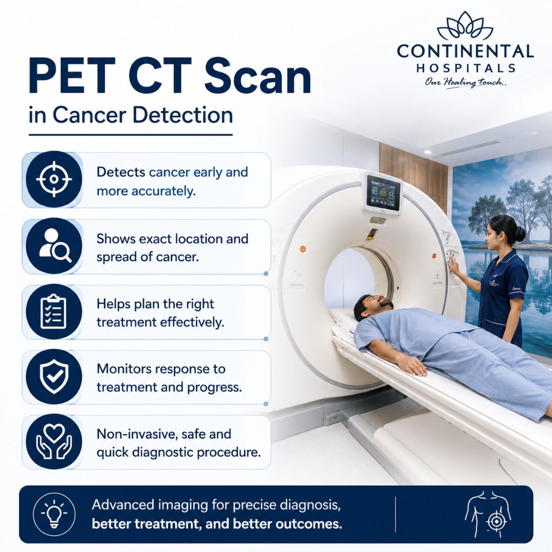
Oncologists rely heavily on a pet scan for cancer management because it provides answers that standard imaging cannot. It serves multiple critical functions throughout a patient's medical journey.

  • Early Phase Detection: It can reveal microscopic cellular changes long before a physical lump or structural change shows up on a standard X-ray.
  • Accurate Disease Staging: It shows whether abnormal cells are confined to one spot or have spread to other areas of the body.
  • Monitoring Treatment Success: It helps doctors see if a tumor is shrinking or becoming less active during chemotherapy or radiation.
  • Spotting Recurrence: It provides early warning signs if abnormal cells begin to regroup after a patient enters remission.

What Should You Expect Inside a PET Scan Machine?

Understanding the physical layout of the test helps ease anxiety. The equipment is designed with patient safety and imaging precision in mind.

  • The Scanner Design: The machine is a large, ring-shaped imaging tunnel with an open-ended design to maximize patient comfort.
  • The Patient Bed: You lie down on a padded, motorized table that slowly guides you through the center of the scanning ring.
  • Quiet Operation: Unlike loud MRI machines, this scanner operates quietly, making the experience calm and stress-free.
  • The Imaging Process: The internal sensors rotate silently inside the housing to capture multi-angle data without moving your body.

How Do You Prepare For the Imaging Procedure?

Proper preparation ensures that your cells absorb the radiotracer correctly, leading to flawless scan results.

  • Fasting Windows: You must fast for at least four to six hours before your appointment, avoiding all food and sugary drinks.
  • Hydration Rules: Drinking plenty of plain, unflavored water is highly encouraged to keep your body hydrated and help flush the tracer out later.
  • Activity Restrictions: Avoid strenuous physical exercise for twenty-four hours prior to the test to prevent muscle tissue from absorbing the tracer.
  • Medical History Review: Inform the nuclear medicine team about all daily medications, especially if you manage diabetes.

Why Choose Continental Hospitals For Your Scan?

Selecting the right diagnostic center is essential for accuracy, safety, and a smooth clinical experience. Continental Hospitals stands out as the premier choice in the region.

Second Opinion

  • Global Accreditations: We are a premier healthcare facility holding prestigious Joint Commission International (JCI) and National Accreditation Board for Hospitals & Healthcare Providers (NABH) accreditations, ensuring world-class safety protocols.
  • Advanced Imaging Infrastructure: Our department features cutting-edge, high-resolution scanners that deliver sharp images for precise diagnoses.
  • Expert Nuclear Medicine Team: Your scans are interpreted by highly experienced, internationally trained nuclear medicine specialists and board-certified radiologists.
  • Safe Healing Environment: As India's first LEED-qualified super specialty hospital building, our infrastructure features natural ventilation and private spaces designed to enhance patient comfort.

Conclusion

A PET CT scan is an invaluable, life-saving resource in modern oncology. By combining detailed anatomical pictures with deep cellular insights, it empowers medical specialists to make fast, accurate, and deeply personalized treatment decisions. For patients navigating a cancer diagnosis, this advanced imaging offers the clarity needed to face the path ahead with confidence.

If you suffer from symptoms that require an expert diagnostic evaluation, or if your primary physician has advised an advanced oncology check, reach out to our dedicated care team. Book a comprehensive consultation with our best medical oncologist in Hyderabad at Continental Hospitals, Hyderabad, to receive world-class care and precise diagnostic insights.

Need an Appointment?

Frequently Asked Questions

A PET CT scan is an advanced imaging test that combines Positron Emission Tomography (PET) and Computed Tomography (CT) into a single examination. The PET component detects metabolic activity in cells by using a small amount of radioactive tracer, usually glucose-based, while the CT component provides detailed anatomical images of organs and tissues. Cancer cells typically consume more glucose than normal cells, making them appear as bright areas on PET images. By combining metabolic and structural information, doctors can identify abnormal tissue more accurately. PET CT scans help detect cancer, determine its extent, evaluate treatment response, and monitor for recurrence. The procedure is noninvasive and provides comprehensive information that may not be available through conventional imaging alone. It has become an essential tool in modern cancer diagnosis and management.
PET CT scans are widely used in cancer detection because they can identify abnormal metabolic activity before significant structural changes become visible. Many cancers show increased glucose uptake, which allows PET imaging to detect suspicious areas throughout the body. The CT component helps pinpoint the exact location of these abnormalities. This combined approach improves diagnostic accuracy and helps physicians distinguish between benign and malignant conditions. PET CT scans are commonly used to detect cancers such as lung cancer, lymphoma, colorectal cancer, breast cancer, and head and neck cancers. They also help determine whether cancer has spread to lymph nodes or distant organs. Early and accurate detection enables timely treatment planning and can significantly improve patient outcomes.
Proper preparation is important to ensure accurate PET CT scan results. Patients are usually advised to avoid eating for several hours before the scan, although drinking water is generally encouraged. Foods high in sugar and strenuous physical activity should be avoided before the test because they can affect glucose metabolism and interfere with image quality. Patients with diabetes may receive specific instructions regarding medications and meal timing. Comfortable clothing should be worn, and metal objects may need to be removed. Inform your doctor about any medications, allergies, pregnancy, or breastfeeding status before the examination. After receiving the tracer injection, patients typically rest quietly for a short period to allow the tracer to distribute throughout the body. Following preparation guidelines helps improve scan accuracy and diagnostic reliability.
During a PET CT scan, a small amount of radioactive tracer is injected into a vein. The tracer travels through the bloodstream and accumulates in tissues based on their metabolic activity. Patients usually wait for approximately 30 to 60 minutes before scanning begins. During the scan, the patient lies on a motorized table that moves through the imaging machine. It is important to remain still to obtain clear images. The scan itself is painless and generally takes between 20 and 45 minutes depending on the area being examined. The PET and CT images are captured during the same session and later combined for detailed analysis. After the procedure, most patients can resume normal activities unless instructed otherwise by their healthcare provider.
Yes, PET CT scans play a major role in cancer staging. Staging refers to determining the size of the tumor and whether cancer has spread to nearby lymph nodes or distant organs. PET CT scans provide detailed information about both the location and metabolic activity of cancerous tissue throughout the body. This comprehensive assessment helps doctors identify the extent of disease more accurately than many conventional imaging methods. Accurate staging is essential for selecting the most appropriate treatment strategy, whether surgery, chemotherapy, radiation therapy, immunotherapy, or a combination of treatments. PET CT scans can also reveal previously undetected metastatic disease, preventing unnecessary procedures and helping healthcare teams develop personalized treatment plans for each patient.
PET CT scans are valuable for evaluating how well cancer treatment is working. Because PET imaging measures metabolic activity, changes in tumor behavior can often be detected before visible changes in tumor size occur. After chemotherapy, radiation therapy, targeted therapy, or immunotherapy, a PET CT scan can show whether cancer cells are becoming less active or responding to treatment. This information helps doctors determine if the current treatment plan is effective or if adjustments are needed. PET CT scans may also identify residual disease after treatment and assist in long term follow up. By providing early insights into treatment response, PET CT imaging supports more informed clinical decisions and improved patient management.
PET CT scans are generally considered safe and are routinely performed worldwide. The radioactive tracer used in the scan contains a very small amount of radiation that typically leaves the body naturally within a short period. The benefits of obtaining accurate diagnostic information usually outweigh the minimal risks associated with radiation exposure. Most patients do not experience side effects from the tracer injection. However, pregnant women and breastfeeding mothers should discuss potential risks with their healthcare provider before undergoing the procedure. Allergic reactions are rare. Patients are often encouraged to drink plenty of fluids after the scan to help eliminate the tracer from the body. Your doctor will recommend the test only when the expected diagnostic benefits are clinically justified.
Yes, PET CT scans are highly effective in detecting cancer recurrence after treatment. Even when patients have completed therapy and appear symptom free, cancer cells may sometimes return. PET CT imaging can identify abnormal metabolic activity suggestive of recurrent disease before it becomes visible on other imaging studies or causes symptoms. Early detection of recurrence allows doctors to intervene quickly and consider additional treatment options. PET CT scans are particularly useful in monitoring cancers that have a higher risk of recurrence. They can also help differentiate between scar tissue from previous treatments and active cancer cells. Regular follow up imaging, when recommended by a healthcare provider, plays an important role in ongoing cancer surveillance and long term patient care.
Disclaimer: The information provided in this blog is intended for general knowledge and informational purposes only, and does not constitute medical advice. Always consult with a qualified healthcare professional for any medical concerns or before making any decisions about your health.

Get in Touch With Us

Whether you are from India or abroad, we are here to help you with your medical queries. Please fill out the form below and our team will connect with you shortly.

  • ✔ Quick response from our medical experts
  • ✔ Secure data handling & privacy
  • ✔ Easy upload for reports & documents
0 / 100
Checkboxes Section


Chat with our medical experts on WhatsApp for quick assistance and hassle-free appointment booking

Recent Posts
Language Based Image
0 / 100